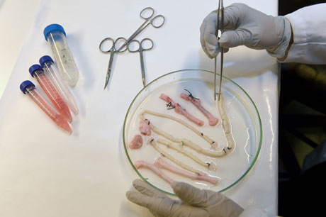
The technology for creating new tissues from stem cells has taken a giant leap forward. Two tablespoons of blood are all that is needed to grow a brand new blood vessel in just seven days. This is shown in a new study from Sahlgrenska Acadedmy and Sahlgrenska University Hospital published in EBioMedicine.
Just three years ago, a patient at Sahlgrenska University Hospital received a blood vessel transplant grown from her own stem cells.
Suchitra Sumitran-Holgersson, Professor of Transplantation Biology at Sahlgrenska Academy, and Michael Olausson, Surgeon/Medical Director of the Transplant Center and Professor at Sahlgrenska Academy, came up with the idea, planned and carried out the procedure.
Missing a vein
Professors Sumitran-Holgersson and Olausson have published a new study in EBioMedicine based on two other transplants that were performed in 2012 at Sahlgrenska University Hospital. The patients, two young children, had the same condition as in the first case – they were missing the vein that goes from the gastrointestinal tract to the liver.
“Once again we used the stem cells of the patients to grow a new blood vessel that would permit the two organs to collaborate properly,” Professor Olausson says.
Stroke of genius
This time, however, Professor Sumitran-Holgersson, found a way to extract stem cells that did not necessitate taking them from the bone marrow.
“Drilling in the bone marrow is very painful,” she says. “It occurred to me that there must be a way to obtain the cells from the blood instead.”
The fact that the patients were so young fueled her passion to look for a new approach. The method involved taking 25 milliliter (approximately 2 tablespoons) of blood, the minimum quantity needed to obtain enough stem cells.
Blood willingly cooperates
Professor Sumitran-Holgersson’s idea turned out to surpass her wildest expectations – the extraction procedure worked perfectly the very first time.
“Not only that, but the blood itself accelerated growth of the new vein,” Professor Sumitran-Holgersson says. “The entire process took only a week, as opposed to a month in the first case. The blood contains substances that naturally promote growth.”
More groups of patients can benefit
Professors Olausson and Sumitran-Holgersson have treated three patients so far. Two of the three patients are still doing well and have veins that are functioning as they should. In the third case the child is under medical surveillance and the outcome is more uncertain.
They researchers have now reached the point that they can avoid taking painful blood marrow samples and complete the entire process in the matter of a week.
“We believe that this technological progress can lead to dissemination of the method for the benefit of additional groups of patients, such as those with varicose veins or myocardial infarction, who need new blood vessels,” Professor Holgersson says. “Our dream is to be able to grow complete organs as a way of overcoming the current shortage from donors.”
The article In vivo application of tissue-engineered veins using autologous peripheral whole blood: A proof of concept study was published in EBioMedicine on 22 October.
Contact:
Suchitra Sumitran-Holgersson, PhD, Professor of Transplantation Biology, Sahlgrenska Academy, Laboratory for Transplantation and Regenerative Medicine, University of Gothenburg, and Department of Surgery, Sahlgrenska University Hospital, phone +46 31-343 0021,
cell +46 72-749 08 08, suchitra.holgersson@gu.se
Michael Olausson, Surgeon/Medical Director, Transplant Center, Sahlgrenska University Hospital, Professor, Sahlgrenska Academy, University of Gothenburg, phone 46 31-342 70 25,
cell 46 70-543 43 60, michael.olausson@surgery.gu.se
Imaging breakthrough. Images generated using the new iDISCO technique show nerves responsible for conducting pain and other sensations, in a mouse embryo (top); axons of the motor nerve that that controls eye movements, in an adult mouse (middle); and collecting ducts that concentrate urine before it leaves the kidney, in an adult mouse (bottom).
Newswise — In a significant technical advance, a team of neuroscientists at The Rockefeller University has devised a fast, inexpensive imaging method for probing the molecular intricacies of large biological samples in three dimensions, an achievement that could have far reaching implications in a wide array of basic biological investigations.
The new method, called iDISCO, optimizes techniques for deep tissue immunolabeling and combines them with recent technological innovations in tissue clearing and light sheet microscopy to achieve unprecedented deep labeling and imaging of molecular structures in the brain, the kidney, and other organs and tissues in experimental settings. A detailed report on iDISCO is in the November 6 issue of the journal Cell.
“What we did was optimize many different parameters of several existing techniques to create this powerful new labeling method,” says Marc Tessier-Lavigne, Rockefeller president, Carson Family Professor, head of the Laboratory of Brain Development and Repair, and senior author on the new study. “These optimization efforts paid off in spades, dramatically extending our ability to visualize molecular structures deep in intact complex tissues like the brain. Although developed in our laboratory to help us pursue neuroscience questions, we believe this new method will provide biological researchers in many disciplines with an important new tool for advancing their work.”
When executed as part of a coordinated protocol, the advance creates a powerful method for imaging molecular structures deep in tissues that is much simpler and more rapid than previous approaches used by biological researchers.
“For us as neuroscientists, one of the big applications of this new imaging method has been the ability to visualize axonal pathways in the developing and adult brain,” says Nicolas Renier, a postdoctoral associate in the Tessier-Lavigne laboratory and co-first author on the study. “We were surprised at just how well we were able to image these detailed structures in the context of the whole brain.”
“The fact that we can now visualize neural circuit formation in larger embryos allows us to study the developing nervous system when it is more well formed,” says Zhuhao Wu, also a postdoctoral associate in the Tessier-Lavigne laboratory and co-first author on the study. “This opens entire new avenues to our research.”
The team focused on three techniques, as they sought to maximize the effectiveness of deep tissue immunolabeling and combine it with innovations by other scientists in tissue clearing and light sheet microscopy.
Clearing is a chemical treatment process that renders biological samples transparent, enabling light to reach deep inside them, and it is in this area particularly that new frontiers have been established in a number of laboratories recently, making it possible to peer deeper into samples than ever before.
Immunolabeling is a longstanding laboratory technique for tagging molecules of interest in biological samples with selected antibodies, but usually in relatively small or thin samples. In this area, the Rockefeller group demonstrated their ability to introduce 28 widely used research antibodies much more deeply and more rapidly into a diversity of samples than had previously been possible. Immunolabeling differs from another commonly used technique in which fluorescent reporter molecules are introduced into a tissue transgenically. Immunolabeling instead attaches tags to endogenous molecules in a study sample.
“Being able to visualize molecules introduced transgenically by the experimenters is a very valuable tool,” says Tessier-Lavigne. “What drove us, however, was a desire to complement that tool with a greater ability to look at endogenous molecular processes. In this area, our new labeling method breaks a barrier we weren’t necessarily expecting to be able to break when we set out on this project.”
Light sheet microscopy has the power to scan whole organs or large tissues in relatively short amounts of time at very high resolutions. The data set captured is so detailed that it can be translated either into images offering a comprehensive view of the entire sample or a closer look at smaller structures within the sample.
Renier and Wu are co-lead authors on the Cell study, and Tessier-Lavigne is senior author. Their coauthors are: David I. Simon, Jing Yang and Pablo Ariel, also in the Tessier-Lavigne laboratory.
A new technique can light up cancer cells so doctors can see them sooner.
Originally published: Nov 11 2014 – 3:00pm
By: Marsha Lewis, Contributing Producer
(Inside Science TV) – Cancer is the second leading cause of death in the United States killing 585,000 Americans every year.
Susann Brady-Kalnay, a cancer biologist at Case Western Reserve University in Cleveland, Ohio wants to change that.
“What happens now with cancer patients is they come in when they have symptoms, and it’s almost too late. The cancer has spread all over their body and caused symptoms,” said Brady-Kalnay.
She wants to catch cancer before symptoms start, with a method called molecular imaging.
“Molecular imaging means that you’re making an imaging agent that actually detects a specific event,” said Brady-Kalnay.
Her team has developed an imaging agent that can be injected into the body. It travels through the bloodstream, finds tumor cells and lights them up so they are visible on MRI scans.
“These probes light up those tumors in two minutes. They travel anywhere in the body,” Brady-Kalnay explained.
In an animation, green areas showed the main tumor mass and the color yellow showed tumor cells that had spread. While performing a procedure, surgeons would not normally be able to see the yellow cancer cells and where they have spread.
“Now, we’re able to allow the surgeon to actually see where the tumor cells are while they’re cutting,” Brady-Kalnay said.
In lab models, the agents detected more than 99% of brain tumor cells. The technique could be used to detect melanoma, prostate and lung cancers sooner.
“That’s our goal here to screen people, to do lifelong screening to find things early because we’ve always known that the best way to treat cancer is to find it early and take it out,” said Brady-Kalnay.
She hopes to begin human clinical trials with her agent in the next two to three years. She says the real key to coming up with this discovery was having a team of chemists, physicists, biomedical engineers and cancer biologists all work collaboratively.
Marsha Lewis is a freelance producer based in California. She has won 11 National Telly Awards and nine Regional Emmy Awards for her work in local and national syndicated news.
Will ultrasound-on-a-chip make medical imaging so cheap that anyone can do it?
By Antonio Regalado on November 3, 2014
Jonathan Rothberg
A scanner the size of an iPhone that you could hold up to a person’s chest and see a vivid, moving, 3-D image of what’s inside is being developed by entrepreneur Jonathan Rothberg.
Rothberg says he has raised $100 million to create a medical imaging device that’s nearly “as cheap as a stethoscope” and will “make doctors 100 times as effective.” The technology, which according to patent documents relies on a new kind of ultrasound chip, could eventually lead to new ways to destroy cancer cells with heat, or deliver information to brain cells.
Rothberg has a knack for marrying semiconductor technology to problems in biology. He started and sold two DNA-sequencing companies, 454 and Ion Torrent Systems (see “The $2 Million Genome” and “A Semiconductor DNA Sequencer”), for more than $500 million. The profits have allowed Rothberg, who showed up for an interview wearing worn chinos and a tattered sailor’s belt, to ply the ocean on a 130-foot yacht named Gene Machine and to indulge high-concept hobbies like sequencing the DNA of mathematical geniuses.
The imaging system is being developed by Butterfly Network, a three-year old company that is the furthest advanced of several ventures that Rothberg says will be coming out of 4Combinator, an incubator he has created to start and finance companies that combine medical sensors with a branch of artificial-intelligence science called deep learning.
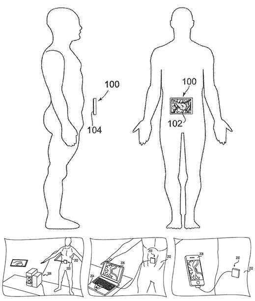
Rothberg won’t say exactly how Butterfly’s device will work, or what it will look like. “The details will come out when we are on stage selling it. That’s in the next 18 months,” he says. But Rothberg guarantees it will be small, cost a few hundred dollars, connect to a phone, and be able to do things like diagnose breast cancer or visualize a fetus.
Butterfly’s patent applications describe its aim as building compact, versatile new ultrasound scanners that can create 3-D images in real time. Hold it up to a person’s chest, and you would look through “what appears to be a window” into the body, according to the documents.
Concept drawings filed with the patent office by Butterfly Network show ideas for a small, 3-D ultrasound imaging device
With the $100 million supplied by Rothberg and investors, which include Stanford University and Germany’s Aeris Capital, Butterfly appears to be placing the largest bet yet by any company on an emerging technology in which ultrasound emitters are etched directly onto a semiconductor wafer, alongside circuits and processors. The devices are known as “capacitive micro-machined ultrasound transducers,” or CMUTs.
Most ultrasound machines use small piezoelectric crystals or ceramics to generate and recieve sound waves. But these have to be carefully wired together, then attached via cables to a separate box to process the signals. Anyone who can integrate ultrasound elements directly onto a computer chip could manufacture them cheaply in large batches, and more easily create the type of arrays needed to produce 3-D images.
“The vision for this product has been around for many years. It remains to be seen whether someone can make it into a market-validated reality.”
Ultrasound is used more often by doctors than any other type of imaging test, including to view a baby during pregnancy, to find tumors in soft tissues like the liver, and more recently to treat prostate cancer by heating up cells with sound waves.
The idea for micromachined ultrasound chips dates to 1994, when Butrus Khuri-Yakub, a Stanford professor who advises Rothberg’s company, built the first one. But none have been a commercial success, despite a decade of interest by companies including General Electric and Philips. This is because they haven’t functioned reliably and have proved difficult to manufacture.
“The vision for this product has been around for many years. It remains to be seen whether someone can make it into a market-validated reality,” says Richard Przybyla, head of circuit design at Chirp Microsystems, a startup in Berkeley, California, that’s developing ultrasound systems that let computers recognize human gestures. “Perhaps what was needed all along is a large investment and a dedicated team.”
Rothberg says he got interested in ultrasound technology because his oldest daughter, now a college student, has tuberous sclerosis. It is a disease that causes seizures and dangerous cysts to grow in the kidneys. In 2011 he underwrote an effort in Cincinnati to test whether high-intensity ultrasound pulses could destroy the kidney tumors by heating them.
What he saw led Rothberg to conclude there was room for improvement. The setup—an MRI machine to see the tumors, and an ultrasound probe to heat them—cost millions of dollars, but wasn’t particularly fast, more like a “laser printer that takes eight days to print and looks like my kids drew it in crayon,” he says. “I set out to make a super-low-cost version of this $6 million machine, to make it 1,000 times cheaper, 1,000 times faster, and a hundred times more precise.”
Rothberg claims there’s a “secret sauce” to Butterfly’s technology, but he won’t reveal it. But it may have as much to do with clever device and circuit design as overcoming the physical limits and manufacturing problems that CMUT technology has faced so far.
One reason to think so is that the company’s cofounder, Nevada Sánchez, previously helped cosmologists design a much cheaper radio telescope with a signal-processing trick called a butterfly network, also the origin of the startup’s name. Also working with the company is Greg Charvat, who joined it from MIT’s Lincoln Laboratory, where he developed radar that can see human bodies even through thick stone walls (see “Seeing like Superman”).
During a visit to 4Combinator’s headquarters, which sits inside a marina in Guilford, Connecticut, Charvat and Sanchez showed off a picture of a penny so detailed you could read the letters and numbers on it. They’d taken the image this spring using a prototype chip. “The ultrasound [industry] is basically back in the 1970s. GE and Siemens are building on old concepts,” says Charvat. With chip manufacturing and a few new ideas from radar, he says, “we can image faster, with a wider field of view, and go from millimeter to micrometer resolution.”
Ultrasound works by shooting out sound and then capturing the echo. It can also create beams of focused energy—and chip-based devices could eventually lead to new systems for killing tumor cells. Small devices might also be used as a way to feed information to the brain (it was recently discovered that that neurons can be activated with ultrasonic waves).
“I think it will become better than a human in saying ‘Does this kid have Down syndrome, or a cleft lip?’ And when people are pressed for time it will be superhuman.”
Rothberg says his first goal will be to market an imaging system cheap enough to be used even in the poorest corners of the world. He says the system will depend heavily on software, including techniques developed by artificial intelligence researchers, to comb through banks of images and extract key features that will automate diagnoses.
“We want it to work like ‘panorama’ on an iPhone,” he says, referring to a smartphone function that steers a picture taker to pan across a vista and automatically assembles a composite image. But in addition to recognizing objects—body parts in the case of a fetal exam—and helping the user locate them, Rothberg says the system would also reach preliminary diagnostic conclusions based on pattern-finding software.
“When I have thousands of these images, I think it will become better than a human in saying ‘Does this kid have Down syndrome, or a cleft lip?’ And when people are pressed for time it will be superhuman,” says Rothberg. “I will make a technician able to do this work.”
Rothberg says his incubator has started three other companies in addition to Butterfly, and he’s given each of them between $5 million and $20 million in seed capital. They include a biotechnology firm, Lam Therapeutics, working on treatments connected to tuberous sclerosis; Hyperfine Research, a startup in stealth mode that hasn’t said what type of technology it is developing; and another company that’s unnamed.
European researchers compare three types of X-ray phase tomography to evaluate which methods perform best for a variety of applications, taking into account both spatial resolution and ability to visualize and quantify relevant features within tissue samples
Released: 18-Oct-2014 9:05 AM EDT
Embargo expired: 21-Oct-2014 11:00 AM EDT
Source Newsroom: American Institute of Physics (AIP)
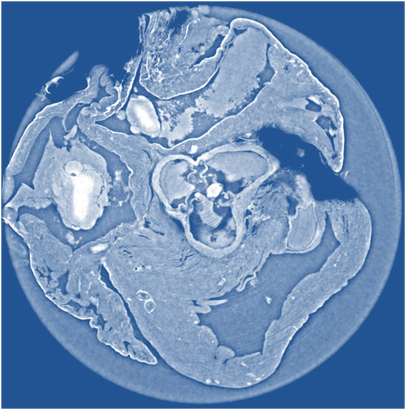
Phase-contrast imaging is a technique for scanning the volumes of soft tissues like tumors or internal organs, but with much greater contrast than conventional CT scans. This image shows a non-invasive “slice” of a rat’s heart tissue made with X-ray phase tomography by propagation-based imaging, which provides sharper data with higher resolution than phase tomography using X-ray grating interferometry (photo:Irene Zanette/Technische Universität München).
Newswise — WASHINGTON, D.C., October 21, 2014 — X-ray phase tomography is an imaging technique that uses penetrating X-rays to create volumetric views through “slices” or sections of soft biological tissues, such as tumors, and it offers strongly enhanced contrast compared to conventional CT scans. Yet scientists still do not know which X-ray phase tomography methods are best suited to yield optimized results for a wide variety of conditions.
To answer this question, a large group of researchers in Europe set out to compare three different X-ray phase tomography methods at the European Synchrotron Radiation Facility’s (ESRF) beamline ID19 in France—X-ray grating interferometry, propagation-based phase tomography with single-distance phase reconstruction, and holotomography.
Led by Irene Zanette, a scientist affiliated with both ESRF and the Technische Universität München (TUM) in Germany, the researchers put these three techniques to the test by examining cancerous tissue from a mouse model and an entire rat’s heart, which they report this week in the Journal of Applied Physics, from AIP Publishing.
Along with colleagues Bert Müller, group leader of the Biomaterials Science Center in Switzerland, and Timm Weitkamp, a scientist at the Synchrotron SOLEIL in France, the team explored which method performs best in terms of spatial resolution and visualization/quantification of relevant features in the samples. They also investigated other related factors such as the simplicity of the setup, and the data acquisition and analysis involved in each method.
To do this, the researchers chose to exploit synchrotron radiation, which produces significantly higher-quality X-rays than conventional X-ray generators such as those found in hospitals.
What exactly is synchrotron radiation? “Think of synchrotron radiation as being analogous to the sort of monochromatic, collimated and intense light produced by lasers, while conventional X-ray generators in hospitals are more analogous to light bulbs we use within our homes,” explained Zanette, currently a postdoctoral scientist in biomedical physics at TUM in Germany.
Importantly, she pointed out, while their study was performed using synchrotron radiation, the same techniques are amenable to both polychromatic and divergent beams and can also be implemented at conventional X-ray sources.
The researchers used an advanced X-ray technique known as “phase-contrast imaging.” This type of imaging works by making the X-ray beam interfere while it propagates from sample to detector, according to Zanette. “This interference is fundamental because it encodes precious information on the phase of the X-ray waves.”
By comparison, conventional X-ray imaging—of the sort performed at hospitals and airports—doesn’t use phase information. Rather, it relies only on the attenuation of the amplitude (reduction in intensity) of the X-ray waves by the object under study to generate image contrast.
“More detailed information is contained in the phase than the amplitude, so it enables us to obtain images with much greater contrast and clearly differentiates cancerous tissue from healthy tissue,” Zanette said.
So what did they find by comparing methods? The group was able to show that for each specimen, the spatial resolution derived from the characteristic morphological features is about twice as good for holotomography and single-distance phase reconstruction compared to X-ray grating interferometry. They also found that X-ray grating interferometry data generally provide much better contrast-to-noise ratios for anatomical features, excel in fidelity of the density measurements, and are more robust against low-frequency artifacts than holotomography.
It turns out that the group regards all three of the phase tomography methods as being complementary. “The application determines which spatial and density resolutions are desired for the imaging task and dose requirements, so it really comes down to a choice between the complexity of the experimental setup and the data processing,” noted Müller. “It’s important to choose the ideal technique for your specific purposes.”
Since synchrotron radiation is of higher quality than the radiation at conventional sources, measurements at synchrotrons represent benchmarking experiments when translating these tomography techniques to clinical practice—especially X-ray grating interferometry, which is attracting attention for use in hospitals.
“Our research should help provide guidance for other researchers in developing an ideal phase-contrast imaging method, which will be adopted by hospitals in the future,” Zanette said.
The article, “Experimental comparison of grating- and propagation-based x-ray phase tomography of soft tissue,” is authored by Sabrina Lang, Irene Zanette, Marco Dominietto, Max Langer, Alexander Rack, Georg Schulz, Geraldine Le Duc, Christian David, Jürgen Mohr, Franz Pfeiffer, Bert Müller, and Timm Weitkamp. It will be published in the Journal of Applied Physics on October 21, 2014 (DOI: 10.1063/1.4897225). After that date, it can be accessed at: http://scitation.aip.org/content/aip/journal/jap/116/15/10.1063/1.4897225
The authors of this paper are affiliated with Technische Universität München in Germany, Biomaterials Science Center in Switzerland, and Synchrotron SOLEIL in France.
ABOUT THE JOURNAL
The Journal of Applied Physics is an influential international journal publishing significant new experimental and theoretical results of applied physics research. See: http://jap.aip.org

 The technology for creating new tissues from stem cells has taken a giant leap forward. Two tablespoons of blood are all that is needed to grow a brand new blood vessel in just seven days. This is shown in a new study from Sahlgrenska Acadedmy and Sahlgrenska University Hospital published in EBioMedicine.
 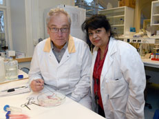 Professors Sumitran-Holgersson and Olausson have published a new study in EBioMedicine based on two other transplants that were performed in 2012 at Sahlgrenska University Hospital. The patients, two young children, had the same condition as in the first case – they were missing the vein that goes from the gastrointestinal tract to the liver.